Teaching AI where to look
Medical-image classification becomes especially difficult when the abnormal region is small, labels are scarce, and much of an image contains anatomy unrelated to the diagnostic question. My research in medical image analysis explores a simple but powerful idea: before asking a model what an image means, it can help to teach it where meaningful anatomy is located.
- Medical Imaging
- Segmentation
- Semi-supervised Learning
- Attention
A radiograph may contain thousands or millions of pixels.
Only a fraction may describe the abnormality of interest.
That creates a fundamental machine-learning problem.
A classifier can technically learn directly from the entire image, but with limited training data it may spend much of its capacity modeling irrelevant structure—or worse, learn shortcuts that correlate with a label without representing the anatomy we actually care about.
Segmentation offers another route.
First identify meaningful regions.
Then use those representations to support the downstream task.
Segment first, classify second
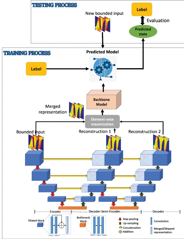
The 2019 IEEE Access study, Regenerative Semi-Supervised Bidirectional W-Network-Based Knee Bone Tumor Classification on Radiographs Guided by Three-Region Bone Segmentation, investigated knee-bone tumor classification from radiographs.
The central idea was not to immediately send the entire radiograph into a classifier.
Instead, the system first separated three major anatomical regions—the femur, tibia, and fibula.
That anatomical information was then incorporated into a broader learning architecture for classifying radiographs as normal, benign, or malignant.
The three-region bone-segmentation component achieved a mean Dice score of 98.06% in the study's evaluation.
When the segmented regions were used for the single-step normal/benign/malignant classification strategy, the reported mean classification accuracy was 85.23%.
These measurements describe different tasks and should not be conflated: Dice evaluates spatial overlap in segmentation, whereas classification accuracy evaluates diagnostic-category predictions.
That distinction is important because a good segmentation does not automatically guarantee a good diagnosis.
It simply gives the downstream model a more focused representation.
Why a W-network?
The architecture also incorporated reconstruction through a bidirectional W-shaped network.
Reconstruction introduces another question during training:
Can the model preserve enough information about the image to recreate meaningful structure?
That task provides an additional source of supervision.
This is particularly relevant when class labels are limited. Medical annotations can be expensive because they may require expert review, while unlabeled or weakly labeled images can be more abundant.
Semi-supervised learning attempts to use that imbalance productively.
Instead of learning only from the final diagnostic labels, the system can also learn structure from the images themselves.
The goal is not to replace clinical labels, but to extract a stronger representation from the available data before using those labels for classification.
Segmentation has its own problem: not every feature deserves to pass through
A second project approaches medical-image analysis from the segmentation side.
The 2020 Applied Sciences study, Enhancing U-Net with spatial-channel attention gate for abnormal tissue segmentation in medical imaging, asks what information should flow through a segmentation network.
U-Net became widely used in biomedical imaging partly because of its skip connections.
As an image moves through the encoder, its representation becomes increasingly abstract. Spatial detail can be lost. Skip connections send higher-resolution information directly from encoder layers to corresponding decoder layers so that the final segmentation can recover precise boundaries.
But there is a catch:
not everything carried by a skip connection is useful.
Where—and what—should the network attend to?
The study introduced a spatial-channel attention gate.
The two parts answer different questions.
Spatial attention: Where in the image is the relevant information?
Channel attention: Which types of learned feature are most useful?
Combining them allows the network to selectively emphasize information before passing encoder features into the decoder.
The method was evaluated across several medical-imaging settings, including polyp, lung-tumor, and brain-tumor segmentation.
On the CVC-ClinicDB evaluation described in the paper, the spatial-channel attention approach achieved a Dice score of 71.72% in the attention-gate comparison, compared with 65.93% for the baseline U-Net.
But the broader contribution is not the six-point difference alone.
It is the architectural idea that a skip connection does not have to be an indiscriminate pipe.
It can itself become a learned decision about which information deserves to move forward.
Segmentation and classification solve different problems
These two projects are closely related, but they should not be presented as the same task.
The knee-radiograph study uses segmentation to guide classification.
The spatial-channel attention study improves segmentation itself.
In the first case, anatomical localization is a step toward predicting a diagnostic category.
In the second, the predicted region is the endpoint being evaluated.
That distinction becomes especially important when reading medical-AI results. A high Dice coefficient, classification accuracy, sensitivity, or AUC each answers a different question.
No single number summarizes the entire system.
| Study direction | Role of regional information | Endpoint to examine |
|---|---|---|
| Segmentation-guided W-network | Bone-region information guides a classification pipeline. | Classification within the radiograph study’s design. |
| Spatial-channel attention U-Net | Attention contributes to a segmentation representation. | Segmentation within the abnormal-tissue study’s design. |
Why this idea still matters
Large foundation models are rapidly changing medical imaging.
Yet the underlying problem has not disappeared.
A model still needs to distinguish relevant anatomy from nuisance information.
It still needs to learn from datasets in which labels may be limited or inconsistent.
And researchers still need to understand whether performance comes from biologically meaningful structures or from shortcuts hidden in the image.
Modern self-supervised and foundation-model approaches offer new tools for representation learning, but segmentation and attention remain valuable precisely because they introduce structure into the learning problem.
They ask the model not merely:
What is this image?
but also:
Which region supports that decision?
That is a much more useful question for the next generation of interpretable medical-imaging systems.
Related publications
Enhancing U-Net with spatial-channel attention gate for abnormal tissue segmentation in medical imaging
Applied Sciences 10.17 (2020), p. 5729 · 2020
Regenerative semi-supervised bidirectional W-network-based knee bone tumor classification on radiographs guided by three-region bone segmentation
IEEE Access 7 (2019), p. 6825-6833 · 2019
See the research in action
Bone tumor detection demo
Watch the demoInterested in this research?
My current interests center on neuroimaging, trustworthy AI, multimodal learning, and foundation models for biomedical data. I am particularly interested in research that connects methodological development with questions of generalizability, interpretation, population differences, and biological relevance.
If your work intersects with these areas—or if you are a student interested in developing research at their intersection—I would be glad to connect.
Get in touch